Home
/ Cow Leg Bones Diagram : Cow Anatomy Posters - In life, this cattle calcaneus is from the right hock and has the smooth side faces outward to the right, as in the photo above.
Cow Leg Bones Diagram : Cow Anatomy Posters - In life, this cattle calcaneus is from the right hock and has the smooth side faces outward to the right, as in the photo above.
Cow Leg Bones Diagram : Cow Anatomy Posters - In life, this cattle calcaneus is from the right hock and has the smooth side faces outward to the right, as in the photo above.. The vertebral column/backbone is the main axis of the skeleton and it protects the spinal cord. Ingredients beef cuts chart and diagram, with photos, names, recipes, and more. Most of the animals have the same bones, although some are shaped differently and placed in different positions. If you dont know the terminologies of the external body parts of a cow and want to know more, you may continue the following part of the article. Baker summary the subsequent discussion is not intended to be a comprehensive review of feet and leg anatomy.
In life, this cattle calcaneus is from the right hock and has the smooth side faces outward to the right, as in the photo above. The cow has a pair of fetlock joints involving the metacarpal bone, the two proximal phalanges and two pairs of sesamoid bones. The pedal bone is the only bone of these three that is completely inside the actual hoof, while the pastern bones serve to connect the hoof to the rest of the leg. Rump, used for steaks, stews and corned beef. It's tough, but when cooked low and slow, it has a nice texture that allows it to shred apart.
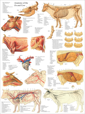
Cow Anatomy Posters from www.dcfirst.com The spinal cord is located in a neural canal formed by a long series of neural arches. The vertebral column/backbone is the main axis of the skeleton and it protects the spinal cord. In the cow the tuber coxae is visible and is readily palpable. General anatomy of the bull and cow ilrated atlas. Cow leg with added bone growth. The cow has a pair of fetlock joints involving the metacarpal bone, the two proximal phalanges and two pairs of sesamoid bones. In types of vertebrae,the neural arch extends as a prominent spine. Thurls need to be wide apart and centrally placed between hip bones and pin bones.
Thurls need to be wide apart and centrally placed between hip bones and pin bones.
The shoulders should not be too straight; Where it is on the cow: Learn all about the most popular beef cuts from our chart, diagram and write up, including popular and alternative names, where the cuts come from on the cow, preferred outdoor cooking methods, their costs relative to each other, and a fantastic recipe for each cut of beef that we've taken from around the web. Figure 3 shows cattle and horse femora. Pastern bone is the fetlock joint and above that the cannon bone of the lower leg. Rump, used for steaks, stews and corned beef. In types of vertebrae,the neural arch extends as a prominent spine. The iliac crest is thin and concave. Of research and development 2 reviewed by g. Ingredients beef cuts chart and diagram, with photos, names, recipes, and more. The tailhead is set slightly above and neatly between pin bones, and the tail is free from coarseness. Cattle and horse long bones also show very distinct differences especially (but not exclusively) femora and metapodials. The ischial tuberosity is triangular in shape.
The bones in the hoof do not entirely formulate the movement of the foot and the leg. In types of vertebrae,the neural arch extends as a prominent spine. The pelvic bones are attached to the back of the spine. Another plan of the both sides of the pelvis in a hanging carcass showing: Anatomy and physiology of domestic animals.
5 Body Parts You Didn T Know You Were There from www.verywellfit.com Clavicle*diagram* clavicle* human*ulnaand*radius* human*humerus* human*humerus*and*scapula. Most of the animals have the same bones, although some are shaped differently and placed in different positions. The aitch bone is curved in steer and bull carcasses, is moderately curved in heifers, but is straight in cow carcasses. General anatomy of the bull and cow ilrated atlas. Only the very bottom of her legs and her two front hooves are visible in this image. Mummified*bear*vs.*human* bear*on*lev* side view of foot bones inter mediate gone gone gone talus gone ca can eug gone cuboid gone gores. The shaft of the bone is then pointing up and back, toward the tail of the animal, to form the distinctive point of the hock in the cow's leg (no. The cranial and caudal dorsal iliac spines.
The bones in the hoof do not entirely formulate the movement of the foot and the leg.
Most of the animals have the same bones, although some are shaped differently and placed in different positions. A) that they shared a common ancestor. Figure 3 shows cattle and horse femora. General anatomy of the bull and cow ilrated atlas. The ischial tuberosity is triangular in shape. Used for choice roasts, the porterhouse and sirloin steaks. The sacral tuber has two prominences; These are covered with horn and often contain dense connective tissue and small nodules of bone. Anatomy and physiology of domestic animals. Cow skeletal anatomy poster 24 x 36 shows the skeleton of the cow with details of the skull, teeth and bones of the legs. Thurls need to be wide apart and centrally placed between hip bones and pin bones. Another plan of the both sides of the pelvis in a hanging carcass showing: In life, this cattle calcaneus is from the right hock and has the smooth side faces outward to the right, as in the photo above.
Mummified*bear*vs.*human* bear*on*lev* side view of foot bones inter mediate gone gone gone talus gone ca can eug gone cuboid gone gores. General anatomy of the bull and cow ilrated atlas. The shoulders should not be too straight; The iliac crest is thin and concave. The pubic bone exposed on a carcass is called the aitch bone.
1 from The skeletal system of a cow by tony smith. A steep shoulder is a good indicator of leg problems. Clavicle*diagram* clavicle* human*ulnaand*radius* human*humerus* human*humerus*and*scapula. Bones of the leg and foot names anatomy functions. This veterinary anatomical atlas includes 27 scientific illustrations with a selection of labelled structures to understand and discover animal anatomy (skeleton, bones, muscles, joints and viscera). If you dont know the terminologies of the external body parts of a cow and want to know more, you may continue the following part of the article. Taken from the area around the breastbone, the brisket is basically the chest or pectoral muscle of the animal. Mummified*bear*vs.*human* bear*on*lev* side view of foot bones inter mediate gone gone gone talus gone ca can eug gone cuboid gone gores.
In types of vertebrae,the neural arch extends as a prominent spine.
Ot 4235 cow femur diagram. The iliac crest is thin and concave. It's tough, but when cooked low and slow, it has a nice texture that allows it to shred apart. 1.5 / 10 ( 2 votes ) skeleton of cow anatomy. The cow has a pair of fetlock joints involving the metacarpal bone, the two proximal phalanges and two pairs of sesamoid bones. Cow leg with added bone growth. This veterinary anatomical atlas includes 27 scientific illustrations with a selection of labelled structures to understand and discover animal anatomy (skeleton, bones, muscles, joints and viscera). The largest and most medial leg bone, forming both the knee and ankle joints. Beef brisket is one of the most flavorful cuts of meat, although it is tough and needs to be cooked in just the right way.it's also a moderately fatty cut of beef, but this can work to your advantage because it tenderizes into succulent, meaty perfection. What does this suggest about mammals? If you dont know the terminologies of the external body parts of a cow and want to know more, you may continue the following part of the article. Used for choice roasts, the porterhouse and sirloin steaks. Pastern bone is the fetlock joint and above that the cannon bone of the lower leg.
/pelvis1500-56aa41da3df78cf772aee25c.jpg)